BACKGROUND
The purpose of this study was to characterize the growth of Campylobacter in broth enrichment (Bolton broth, brain heart infusion broth), material cecum (in vitro), and in broiler live naturally infected (in vivo) in terms of lag period the mean and time generation and The maximum growth rate and population (cell concentration) is reached.
METHOD
Bolton and brain heart infusion broth and recovered materials cecum poultry inoculated with 10 strains of Campylobacter (eight Campylobacter jejuni and Campylobacter coli two), incubated in microaerobic conditions, and the concentration of Campylobacter determined periodically using ISO 10 272: 2006. Methods caeca than 10 flock, which infected in the first thinning, is used to characterize the growth of Campylobacter in live birds. mean generation time (G) (initial lag phase to exponential) is calculated using the formula: G = t / 3.3 logb / B. Mean time lag and μmax calculated using Micro Fit (©) Software (Version 1.0, the Institute of Food Research). Statistical comparison is done by using GENSTAT ver. 14.1 (VSN International Ltd., Hemel Hempstead, UK).
RESULTS
Lag period meant Bolton broth, brain heart infusion broth, cecum material, and the live bird was estimated to be 6.6, 6.7, 12.6, and 31.3 hours, respectively. The corresponding mean generation times of 2.1, 2.2, 3.1, and 6.7 hours, respectively; The maximum growth rate was 0.7, 0.8, 0.4, and 2 generations h (-1) and the maximum population obtained in each matrix were 9.6, 9.9, 7.8, and 7.4 log10 CFU / g, respectively.
CONCLUSION
This study provides data on the growth of Campylobacter in various media lab, the contents of the cecum, and broiler can be used to develop predictive models and / or inform the control strategy based on science as the maximum time between testing herd and slaughter, slaughter logistics, and one stage depopulation unit broiler.
Analysis of 11 years of microbial keratitis in the South West of England using a brain-heart infusion broth.
The purpose of this study was to identify the organisms responsible for microbial keratitis, as identified by the friction of the cornea using a brain-heart infusion broth, trends over time and antimicrobial susceptibility, over a period of 11 years in the two units of the South West of England; Bristol Eye Hospital and the Royal United Hospital, corneal scratches Bath.All done and sent for microbiological analysis between 4 April 2006 and October 31, 2017 in the two units of the retrospective review.
The first line treatment is monotherapy with levofloxacin 0.5% and second-line treatment is a combination of cefuroxime 5% and 1.5% gentamicin. Both direct and enriching culture used.In total, 2614 scratch the cornea of 2116 patients (1082 women, average age 47.7 ± 21.2 years) were identified. 38.1% (n = 996) were positive cultures and organism cultured in 1195. In all, 91.6% is bacteria (69.4% were gram-positive, gram negative 30.6%).
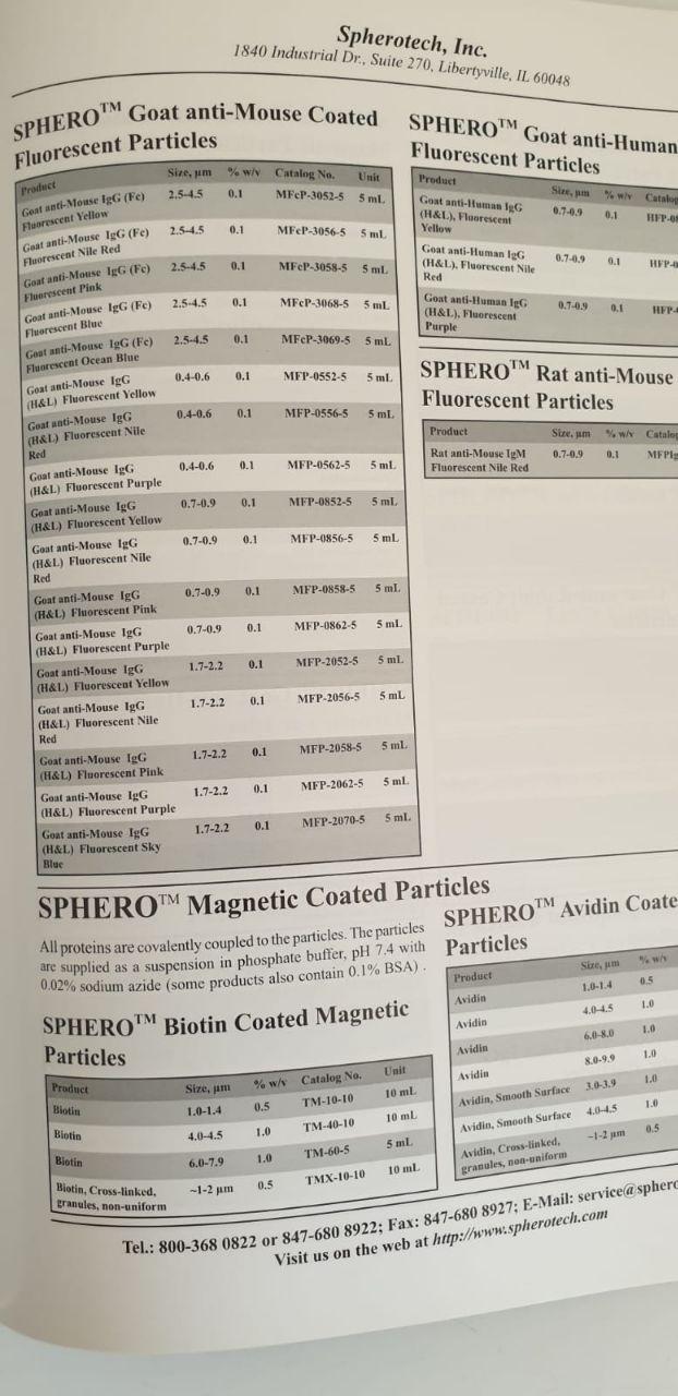
 Human Resistin Antibody |
|
32138-05111 |
AssayPro |
150 ug |
EUR 215 |
Human Resistin Antibody |
|
GWB-F7AAE4 |
GenWay Biotech |
0.1 mg |
Ask for price |
 (Human) - Antibody) Resistin (43-65) (Human) - Antibody |
|
H-028-39 |
PHOENIX PEPTIDE |
50 μl |
EUR 604.8 |
 (Human) - Antibody) Resistin (51-108) (Human) - Antibody |
|
H-028-40 |
PHOENIX PEPTIDE |
50 μl |
EUR 604.8 |
 Anti-Human Resistin Antibody |
|
102-P92G |
ReliaTech |
100 µg |
EUR 245.7 |
|
Description: Resistin belongs to a family of tissue-specific cytokines termed FIZZ (found in inflammatory zones) and RELM. The three known members of this family; Resistin, RELM-α and RELM-β share a highly conserved C-terminal domain, characterized by 10 cysteine residues with a unique spacing motif of C-X11-C-X8-C-X-C-X3-C-X10-C-X-C-X-C-X9-C-C. Resistin is an adipose-derived cytokine (adipokine) whose physiological function and molecular targets are largely unknown. Studies have shown that Resistin suppresses insulin's ability to stimulate glucose uptake, and postulated that Resistin might be an important link between obesity and Type 2 diabetes. Other studies have indicated that Resistin expression is severely suppressed in obesity and that it may act as a feedback regulator of Adipogenesis. |
Anti-Human Resistin Antibody |
|
101-M170 |
ReliaTech |
500 µg |
EUR 246.75 |
|
Description: Resistin belongs to a family of tissue-specific cytokines termed FIZZ (found in inflammatory zones) and RELM. The three known members of this family; Resistin, RELM-α and RELM-β share a highly conserved C-terminal domain, characterized by 10 cysteine residues with a unique spacing motif of C-X11-C-X8-C-X-C-X3-C-X10-C-X-C-X-C-X9-C-C. Resistin is an adipose-derived cytokine (adipokine) whose physiological function and molecular targets are largely unknown. Studies have shown that Resistin suppresses insulin's ability to stimulate glucose uptake, and postulated that Resistin might be an important link between obesity and Type 2 diabetes. Other studies have indicated that Resistin expression is severely suppressed in obesity and that it may act as a feedback regulator of Adipogenesis. Recombinant human Resistin is a 19.5 kDa disulfide-linked homodimeric protein composed of two identical 92 amino acid chains linked by a single disulfide bond. |
Anti-Human Resistin Antibody |
|
101-M618 |
ReliaTech |
100 µg |
EUR 399 |
|
Description: Resistin belongs to a family of tissue-specific cytokines termed FIZZ (found in inflammatory zones) and RELM. The three known members of this family; Resistin, RELM-α and RELM-β share a highly conserved C-terminal domain, characterized by 10 cysteine residues with a unique spacing motif of C-X11-C-X8-C-X-C-X3-C-X10-C-X-C-X-C-X9-C-C. Resistin is an adipose-derived cytokine (adipokine) whose physiological function and molecular targets are largely unknown. Studies have shown that Resistin suppresses insulin's ability to stimulate glucose uptake, and postulated that Resistin might be an important link between obesity and Type 2 diabetes. Other studies have indicated that Resistin expression is severely suppressed in obesity and that it may act as a feedback regulator of Adipogenesis. |
) Human Resistin (RETN) |
|
1-CSB-EP019573HU |
Cusabio |
-
Ask for price
-
Ask for price
-
Ask for price
-
Ask for price
-
Ask for price
-
Ask for price
|
- 100ug
- 10ug
- 1MG
- 200ug
- 500ug
- 50ug
|
|
|
|
Description: Recombinant Human Resistin(RETN) expressed in E.coli |
 Biotinylated Anti-Human Resistin |
|
GWB-BIG87D |
GenWay Biotech |
1 kit |
Ask for price |
 RECOMBINANT HUMAN RESISTIN |
|
GWB-F4A137 |
GenWay Biotech |
0.02 mg |
Ask for price |
 Recombinant Human Resistin |
|
cyt-456-1mg |
ProSpec Tany |
1mg |
EUR 4680 |
Recombinant Human Resistin |
|
cyt-456-25g |
ProSpec Tany |
25µg |
EUR 145 |
Recombinant Human Resistin |
|
cyt-456-5g |
ProSpec Tany |
5µg |
EUR 60 |
 (Human) - RIA Kit) Resistin (43-65) (Human) - RIA Kit |
|
RK-028-39 |
PHOENIX PEPTIDE |
125 tubes |
EUR 1061.64 |
 human Resistin ELISA KIT |
|
201-12-0339 |
SunredBio |
96 tests |
EUR 528 |
|
|
|
Description: A quantitative ELISA kit for measuring Human in samples from biological fluids. |
 Human Resistin ELISA kit |
|
E01A7519 |
BlueGene |
96T |
EUR 700 |
|
Description: ELISA |
Human Resistin ELISA kit |
|
E01R0351-192T |
BlueGene |
192 tests |
EUR 1524 |
|
|
|
Description: A competitive ELISA for quantitative measurement of Human Resistin in samples from blood, plasma, serum, cell culture supernatant and other biological fluids. This is a high quality ELISA kit developped for optimal performance with samples from the particular species. |
Human Resistin ELISA kit |
|
E01R0351-48 |
BlueGene |
1 plate of 48 wells |
EUR 624 |
|
|
|
Description: A competitive ELISA for quantitative measurement of Human Resistin in samples from blood, plasma, serum, cell culture supernatant and other biological fluids. This is a high quality ELISA kit developped for optimal performance with samples from the particular species. |
Human Resistin ELISA kit |
|
E01R0351-96 |
BlueGene |
1 plate of 96 wells |
EUR 822 |
|
|
|
Description: A competitive ELISA for quantitative measurement of Human Resistin in samples from blood, plasma, serum, cell culture supernatant and other biological fluids. This is a high quality ELISA kit developped for optimal performance with samples from the particular species. |
 Human Resistin ELISA Kit |
|
E16HE0035 |
EnoGene |
96T |
EUR 833.33 |
 Human Resistin ELISA KIT |
|
E16HE0035-048 |
EnoGene |
48 wells |
EUR 295 |
Human Resistin ELISA KIT |
|
E16HE0035-096 |
EnoGene |
96 wells |
EUR 395 |
Human Resistin ELISA kit |
|
E22-HC136.48 |
EnoGene |
48T |
EUR 295 |
Human Resistin ELISA kit |
|
E22-HC136.96 |
EnoGene |
96T |
EUR 395 |
Human Resistin ELISA Kit |
|
E28L0045 |
EnoGene |
96T |
EUR 666.67 |
Human Resistin ELISA KIT |
|
E42EH-93 |
EnoGene |
96T/48T |
Ask for price |
Human Resistin ELISA Kit |
|
EK2351 |
SAB |
96 tests |
EUR 497 |
|
|
Human Resistin ELISA Kit |
|
EHR0358 |
Abclonal |
96Tests |
EUR 625.2 |
Human Resistin ELISA KIT |
|
CSB-E06884h-24T |
Cusabio |
1 plate of 24 wells |
EUR 198 |
|
|
|
Description: Quantitativesandwich ELISA kit for measuring Human Resistin in samples from serum, plasma, tissue homogenates. A new trial version of the kit, which allows you to test the kit in your application at a reasonable price. |
Human Resistin ELISA KIT |
|
1-CSB-E06884h |
Cusabio |
-
Ask for price
-
Ask for price
-
Ask for price
|
- 1 plate of 96 wells
- 10 plates of 96 wells each
- 5 plates of 96 wells each
|
|
|
|
Description: Quantitativesandwich ELISA kit for measuring Human Resistin in samples from serum, plasma, tissue homogenates. Now available in a cost efficient pack of 5 plates of 96 wells each, conveniently packed along with the other reagents in 5 separate kits. |
Human Resistin ELISA kit |
|
55R-1714 |
Fitzgerald |
1 kit |
EUR 717 |
|
|
|
Description: ELISA kit for the detection of Human Resistin in the research laboratory |
Human Resistin ELISA kit |
|
55R-2127 |
Fitzgerald |
96 tests |
EUR 825 |
|
Description: ELISA Kit for detection of Resistin in the research laboratory |
Human Resistin ELISA kit |
|
55R-E50 |
Fitzgerald |
96 wells |
EUR 715 |
|
Description: ELISA kit for the detection of Human Resistin in the research laboratory |
human Resistin ELISA KIT |
|
CN-04165H1 |
ChemNorm |
96T |
EUR 530.4 |
human Resistin ELISA KIT |
|
CN-04165H2 |
ChemNorm |
48T |
EUR 351.6 |
Human Resistin ELISA kit |
|
LF-EK50441 |
Abfrontier |
1×96T |
EUR 790.8 |
Human Resistin ELISA Kit |
|
LF-EK70021 |
Abfrontier |
1×96T |
EUR 625.2 |
Human Resistin ELISA Kit |
|
RK00139 |
Abclonal |
96 Tests |
EUR 280 |
Human Resistin ELISA Kit |
|
GWB-SKR059 |
GenWay Biotech |
96 Tests |
Ask for price |
Human Resistin ELISA Kit |
|
GWB-ZZD207 |
GenWay Biotech |
1x96 well plate |
Ask for price |
Human Resistin ELISA Kit |
|
SL1530Hu |
Sunlong |
96 Tests |
EUR 468 |
|
|
 Anti- Resistin Mouse Antibody |
|
GWB-11F9B7 |
GenWay Biotech |
0.1 mg |
Ask for price |
|
|
 Resistin, human recombinant |
|
4559-100 |
Biovision |
each |
EUR 874.8 |
Resistin, human recombinant |
|
4559-1000 |
Biovision |
each |
EUR 6127.2 |
Resistin, human recombinant |
|
4559-25 |
Biovision |
each |
EUR 307.2 |
 Human) Resistin (Recombinant) Human |
|
GWB-8EE40A |
GenWay Biotech |
0.025 mg |
Ask for price |
 Mouse Monoclonal anti-human Resistin |
|
hAP-0528 |
Angio Proteomie |
100ug |
EUR 250 |
 Anti-Mouse Resistin Antibody |
|
103-M461 |
ReliaTech |
100 µg |
EUR 399 |
|
Description: Resistin belongs to a family of tissue-specific cytokines termed FIZZ (found in inflammatory zones) and RELM. The three known members of this family; Resistin, RELM-α and RELM-β share a highly conserved C-terminal domain, characterized by 10 cysteine residues with a unique spacing motif of C-X11-C-X8-C-X-C-X3-C-X10-C-X-C-X-C-X9-C-C. Resistin is an adipose-derived cytokine (adipokine) whose physiological function and molecular targets are largely unknown. Studies have shown that Resistin suppresses insulin's ability to stimulate glucose uptake, and postulated that Resistin might be an important link between obesity and Type 2 diabetes. Other studies have indicated that Resistin expression is severely suppressed in obesity and that it may act as a feedback regulator of Adipogenesis. Recombinant murine Resistin is a 20.2 kDa disulfide-linked homodimeric protein composed of two identical 94 amino acid chains linked by a single disulfide bond. |
Anti-Mouse Resistin Antibody |
|
103-P51G |
ReliaTech |
100 µg |
EUR 245.7 |
|
Description: Resistin belongs to a family of tissue-specific cytokines termed FIZZ (found in inflammatory zones) and RELM. The three known members of this family; Resistin, RELM-α and RELM-β share a highly conserved C-terminal domain, characterized by 10 cysteine residues with a unique spacing motif of C-X11-C-X8-C-X-C-X3-C-X10-C-X-C-X-C-X9-C-C. Resistin is an adipose-derived cytokine (adipokine) whose physiological function and molecular targets are largely unknown. Studies have shown that Resistin suppresses insulin's ability to stimulate glucose uptake, and postulated that Resistin might be an important link between obesity and Type 2 diabetes. Other studies have indicated that Resistin expression is severely suppressed in obesity and that it may act as a feedback regulator of Adipogenesis. Recombinant murine Resistin is a 20.2 kDa disulfide-linked homodimeric protein composed of two identical 94 amino acid chains linked by a single disulfide bond. |
 Protein) Human Resistin (RETN) Protein |
|
20-abx651562 |
Abbexa |
-
Ask for price
-
Ask for price
-
Ask for price
-
Ask for price
-
Ask for price
|
- 100 ug
- 10 ug
- 1 mg
- 200 ug
- 50 ug
|
|
|
Human Resistin (RETN) Protein |
|
abx670217-25ug |
Abbexa |
25 ug |
EUR 661.2 |
|
|
Human Resistin (RETN) Protein |
|
20-abx068871 |
Abbexa |
-
Ask for price
-
Ask for price
-
Ask for price
-
Ask for price
-
Ask for price
|
- 100 ug
- 10 ug
- 1 mg
- 200 ug
- 50 ug
|
|
|
Human Resistin (RETN) Protein |
|
abx262050-25mg |
Abbexa |
2.5 mg |
EUR 325 |
Human Resistin (RETN) Protein |
|
abx262253-10mg |
Abbexa |
10 mg |
EUR 325 |
Human Resistin (RETN) Protein |
|
abx262253-25mg |
Abbexa |
25 mg |
EUR 5975 |
Human Resistin (RETN) Protein |
|
abx262253-5mg |
Abbexa |
5 mg |
EUR 225 |
Human Resistin (RETN) Protein |
|
abx068871-10g |
Abbexa |
10 µg |
EUR 375 |
Human Resistin (RETN) Protein |
|
abx068871-2g |
Abbexa |
2 µg |
EUR 212.5 |
Human Resistin (RETN) Protein |
|
abx670217-50g |
Abbexa |
50 µg |
EUR 462.5 |
Human Resistin (RETN) Protein |
|
abx651562-100g |
Abbexa |
100 µg |
EUR 1062.5 |
Human Resistin (RETN) Protein |
|
abx651562-10g |
Abbexa |
10 µg |
EUR 587.5 |
Human Resistin (RETN) Protein |
|
abx651562-50g |
Abbexa |
50 µg |
EUR 875 |
Human Resistin (RETN) Protein |
|
abx682081-100g |
Abbexa |
100 µg |
EUR 812.5 |
Human Resistin (RETN) Protein |
|
abx682081-20g |
Abbexa |
20 µg |
EUR 312.5 |
 (Human) - FAM Labeled) Resistin (43-65) (Human) - FAM Labeled |
|
FG-028-39A |
PHOENIX PEPTIDE |
1 nmol |
EUR 604.8 |
 (Human) - Purified IgG Antibody) Resistin (51-108) (Human) - Purified IgG Antibody |
|
G-028-40 |
PHOENIX PEPTIDE |
200 μg |
EUR 604.8 |
 Polyclonal Anti- Resistin Antibody |
|
GWB-BBP350 |
GenWay Biotech |
0.1 mg |
Ask for price |
 Recombinant Human Resistin, HEK |
|
cyt-1183-10g |
ProSpec Tany |
10µg |
EUR 145 |
Recombinant Human Resistin, HEK |
|
cyt-1183-1mg |
ProSpec Tany |
1mg |
EUR 5200 |
Recombinant Human Resistin, HEK |
|
cyt-1183-2g |
ProSpec Tany |
2µg |
EUR 60 |
) Recombinant Human Resistin (RETN) |
|
CSB-EP019573HU |
Cusabio |
2058 mg |
Ask for price |
) Recombinant Human Resistin(RETN) |
|
AP70712 |
SAB |
1mg |
EUR 1978 |
|
|
Recombinant Human Resistin (RETN) |
|
Z100805 |
ABM |
25 µg |
EUR 85 |
|
Description: Resistin was identified by screening for genes that are induced during the differentiation of adipocytes but down-regulated in mature adipocytes. Resistin gene expression is induced during the differentiation of adipocytes. Resistin circulates in mouse serum, and its level is increased markedly in obesity. Adipocytes secrete resistin as a unique signaling molecule that may be the hormone potentially linking obesity to type II diabetes, which is characterized by target-tissue resistance to insulin. TNF-alpha is a negative regulator of Resistin gene expression and inhibits Resistin mRNA expression and protein secretion by 70-90% in 3T3-L1 adipocytes. |
Recombinant Human Resistin (RETN) |
|
Z100809 |
ABM |
1.0 mg |
EUR 1500 |
|
Description: Resistin was identified by screening for genes that are induced during the differentiation of adipocytes but down-regulated in mature adipocytes. Resistin gene expression is induced during the differentiation of adipocytes. Resistin circulates in mouse serum, and its level is increased markedly in obesity. Adipocytes secrete resistin as a unique signaling molecule that may be the hormone potentially linking obesity to type II diabetes, which is characterized by target-tissue resistance to insulin. TNF-alpha is a negative regulator of Resistin gene expression and inhibits Resistin mRNA expression and protein secretion by 70-90% in 3T3-L1 adipocytes. |
 CLIA Kit) Human Resistin (RETN) CLIA Kit |
|
20-abx492128 |
Abbexa |
-
Ask for price
-
Ask for price
-
Ask for price
|
- 10 × 96 tests
- 5 × 96 tests
- 96 tests
|
|
|
Human Resistin (RETN) CLIA Kit |
|
abx195057-96tests |
Abbexa |
96 tests |
EUR 990 |
|
|
 ELISA kit for Human Resistin |
|
KET6029-48T |
Abbkine |
48T |
EUR 242.4 |
|
|
|
Description: Quantitative sandwich ELISA for measuring Human Resistin in samples from cell culture supernatants, serum, whole blood, plasma and other biological fluids. |
ELISA kit for Human Resistin |
|
KET6029-5platesof96wells |
Abbkine |
5 plates of 96 wells |
EUR 1292.4 |
|
|
|
Description: Quantitative sandwich ELISA for measuring Human Resistin in samples from cell culture supernatants, serum, whole blood, plasma and other biological fluids. |
ELISA kit for Human Resistin |
|
KET6029-96T |
Abbkine |
96T |
EUR 346.8 |
|
|
|
Description: Quantitative sandwich ELISA for measuring Human Resistin in samples from cell culture supernatants, serum, whole blood, plasma and other biological fluids. |
 Resistin |
|
cyt-456 |
ProSpec Tany |
5µg |
EUR 60 |
|
Description: Recombinant Human Resistin |
Resistin |
|
AP89468 |
SAB |
1mg |
EUR 2640 |
|
|
Resistin |
|
AP78694 |
SAB |
1mg |
EUR 2640 |
|
|
Resistin |
|
AP78695 |
SAB |
1mg |
EUR 2640 |
|
|
 ELISA Kit) Human Resistin (RETN) ELISA Kit |
|
DLR-RETN-Hu |
DL Develop |
96T |
EUR 380 |
|
|
|
Description: serum, plasma, tissue homogenates, cell lysates, cell culture supernates or other biological fluids. |
Human Resistin (RETN) ELISA Kit |
|
DLR-RETN-Hu-48T |
DL Develop |
48T |
EUR 529.2 |
|
|
|
Description: A sandwich quantitative ELISA assay kit for detection of Human Resistin (RETN) in samples from serum, plasma, tissue homogenates, cell lysates, cell culture supernates or other biological fluids. |
Human Resistin (RETN) ELISA Kit |
|
DLR-RETN-Hu-96T |
DL Develop |
96T |
EUR 684 |
|
|
|
Description: A sandwich quantitative ELISA assay kit for detection of Human Resistin (RETN) in samples from serum, plasma, tissue homogenates, cell lysates, cell culture supernates or other biological fluids. |
Coagulase-negative Staphylococcus (cons) is the most common organism cultured (n = 430). Pseudomonas aeruginosa is a gram-negative organisms most frequently identified (n = 189). In total, 6.9% (n = 83) cultured organism is a fungus. In all, 1.4% (n = 17) is Acanthamoeba. No statistically significant trend in the organism were observed during the study period. sensitivity testing confirmed reasonable sensitivity towards empirical antibiotics used in clinical practice.This is the first report on the trend of microbial keratitis in the South West of England. virulent organisms that may be detected in the culture directly, whereas a low virulent organisms such as the cons are more likely to be detected in enrichment only. Antibiotic sensitivity testing confirmed fluoroquinolone monotherapy as first-line treatment accordingly.